Diagnostic Hysteroscopy: Risks, How to Prepare & What to Expect

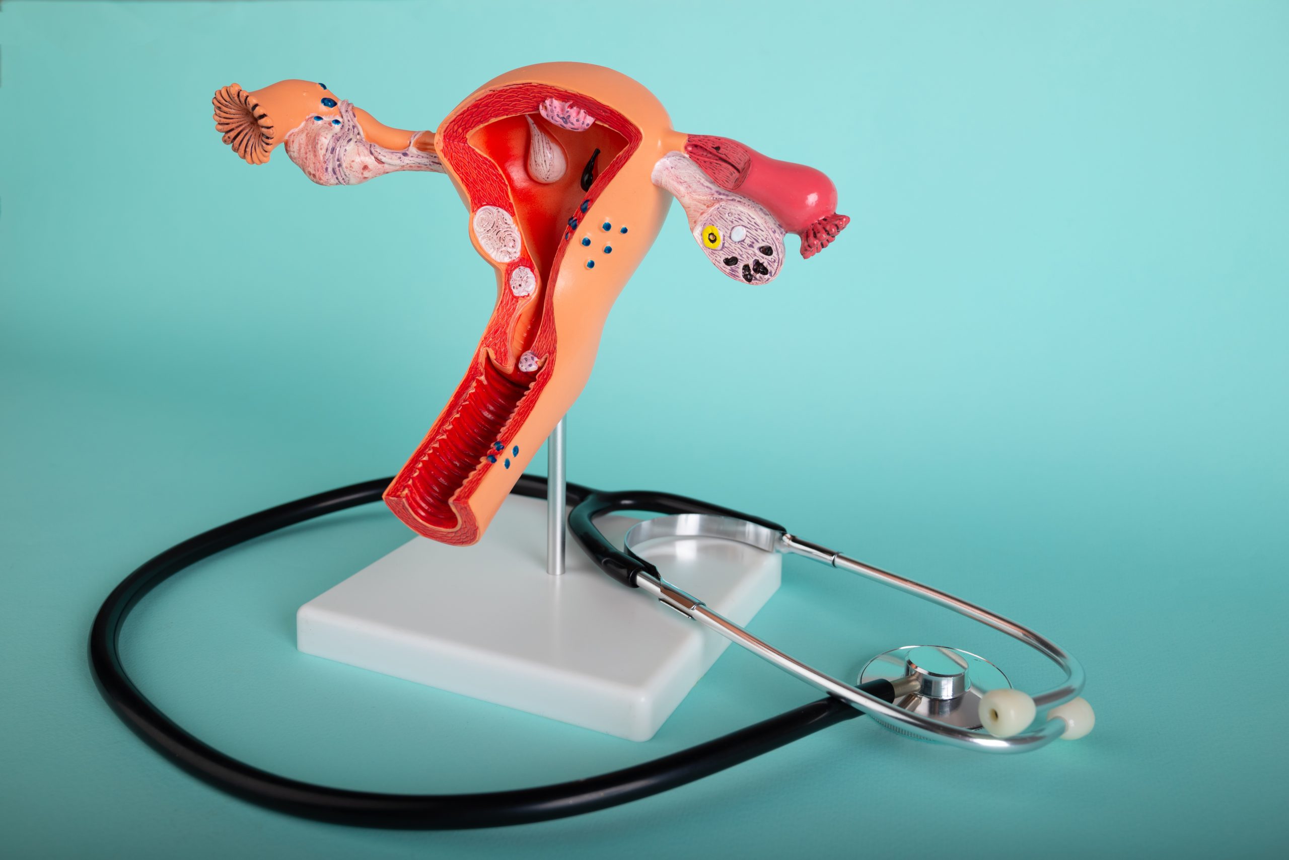
A diagnostic hysteroscopy is a quick, minimally invasive procedure that lets your provider look directly inside the uterus using a thin, lighted camera called a hysteroscope.
The camera is gently passed through the vagina and cervix without any incisions or stitches. Sterile saline is then used to expand the uterus, allowing a clear view of the uterine walls and lining. The entire process usually takes less than 10 minutes.
Keep in mind that this is a great, minimally invasive approach to diagnosis, but you will likely need an operative hysteroscopy or appropriate procedure to resolve any diagnosis.
Why Is a Diagnostic Hysteroscopy Performed?
A hysteroscopy is one of the most accurate ways to assess the uterine lining, also called the endometrium, and identify structural issues that may affect fertility or menstrual health. It can help diagnose :
-
Uterine polyps
-
Fibroids
-
Scar tissue, also called adhesions
-
Congenital abnormalities
-
Other structural or pathological conditions affecting the uterine cavity
This type of evaluation is especially helpful for people experiencing infertility, abnormal uterine bleeding, or recurrent pregnancy loss. It is also commonly performed prior to fertility treatment such as IVF.
When Is a Diagnostic Hysteroscopy Scheduled?
For the clearest view of the uterine cavity, a diagnostic hysteroscopy is typically performed:
- After menstrual bleeding has ended
- Before ovulation, usually between cycle days 6 and 12
Your provider will help determine the best timing based on your cycle and treatment plan.
How to Prepare
Your provider may give you specific instructions, but preparation for a diagnostic hysteroscopy often includes:
- Taking 800 mg of ibuprofen about one hour before your appointment to help reduce cramping
- Eating and drinking normally since there are no dietary restrictions
- Arriving 30 minutes early to allow time for check-in
- Emptying your bladder before the procedure
- Completing a pregnancy test before the procedure begins
Your care team may adjust these instructions based on your health and treatment plan.
What Happens During the Procedure
The exact steps may vary slightly depending on your provider and your individual needs. A diagnostic hysteroscopy generally includes:
- Gently inserting a speculum to visualize the cervix
- Passing a thin, lighted hysteroscope through the cervix and into the uterus
- Filling the uterus with sterile saline to improve visibility
- Viewing the uterine cavity on a monitor, with the option to watch if you prefer
- Completing the evaluation within just a few minutes
Most patients describe the sensation as mild to moderate cramping, similar to menstrual cramps.
Results After Your Hysteroscopy
Once the procedure is completed, your provider will review the findings with you. You may have been able to watch the hysteroscopy in real time on a monitor. Results generally fall into two categories:
- Normal findings. No abnormalities were seen inside the uterus.
- Abnormal findings. These may include polyps, fibroids, scar tissue, congenital differences in the uterine shape, or areas of abnormal bleeding.
If anything abnormal is identified, your provider will discuss the next steps, which may include:
- Additional imaging
- Removal of a polyp or fibroid
- Treatment of adhesions
- Planning an operative hysteroscopy if needed
- Adjustments to your fertility treatment plan
What Are the Risks of a Hysteroscopy?
Complications from a diagnostic hysteroscopy are rare and occur in about 3 to 6 percent of cases. Potential risks include:
- Fluid overload caused by excess fluid absorption
- Electrolyte imbalance, meaning changes in the body’s salt and water levels
- Rare fluid accumulation in the lungs or brain
- Uterine irritation or, rarely, perforation
- Infection
Your provider will review these risks with you and answer any questions you may have.
What to Expect After the Procedure
Recovery is usually quick, and most patients resume normal activities the same day. It is common to experience:
- Light to moderate cramping
- Spotting or light bleeding for up to one week
- Watery discharge as the saline drains
You can generally take ibuprofen as needed for comfort and should use peri-pads rather than tampons during this time. Avoid sexual intercourse for one week after the procedure, and always follow the specific instructions given by your medical provider and care team.
When to Call the Office
Contact your provider right away if you experience:
- Fever or chills
- Heavy bleeding such as soaking more than one maxi pad per hour
- Severe abdominal pain not relieved by ibuprofen
- Severe nausea, vomiting, or headache
These symptoms may indicate a complication and should be evaluated promptly.
Who Should Not Have a Diagnostic Hysteroscopy
Patients who should not have a diagnostic hysteroscopy include those with :
- known uterine anomalies that should be surgically resected
- cervical cancer
- viable uterine pregnancy
The Bottom Line
A diagnostic hysteroscopy is a short, simple procedure that offers valuable insight into the health of the uterus. It is one of the most effective ways to diagnose polyps, fibroids, scar tissue, and other issues that may affect fertility or menstrual health.
Most people tolerate it well, recover quickly, and benefit from the clear information it provides. Whether your results are normal or show something that needs treatment, a hysteroscopy helps guide the next steps in your reproductive care with clarity and confidence.